

Advanced Retinal & Optic Nerve Imaging
Documented, comparable, and completed in a single visit
The single most useful thing an eye exam can produce is a record precise enough to compare against next year. Subtle change is what matters in glaucoma, macular disease, diabetic retinopathy, and corneal conditions, and the human eye examining another human eye is not a reliable instrument for detecting it.
OPTOS ultra widefield imaging photographs a large portion of the retina in a single capture, frequently including peripheral areas where retinal tears, diabetic change, and pigmented lesions appear first. Maestro OCT resolves the individual retinal layers to a fine level of detail, and OCT angiography maps blood flow through the retinal capillaries without dye.
Antares corneal topography maps the corneal surface for keratoconus screening, specialty lens fitting, and refractive surgery planning.
What the imaging can reveal
Retinal tears and detachments, macular degeneration, epiretinal membranes, macular holes, diabetic retinopathy, vascular occlusions, optic nerve changes from glaucoma, choroidal nevi, and corneal irregularity are all detectable and measurable with this equipment. Many of these produce no symptoms at all in their early stages.
Systemic disease also leaves signatures in the retina. Hypertension, elevated cholesterol, and certain neurological conditions can produce findings that warrant a conversation with your physician.
Imaging is retained in your record, so each subsequent visit compares against your own prior scans rather than against a population average. That comparison is where the real diagnostic value lives.
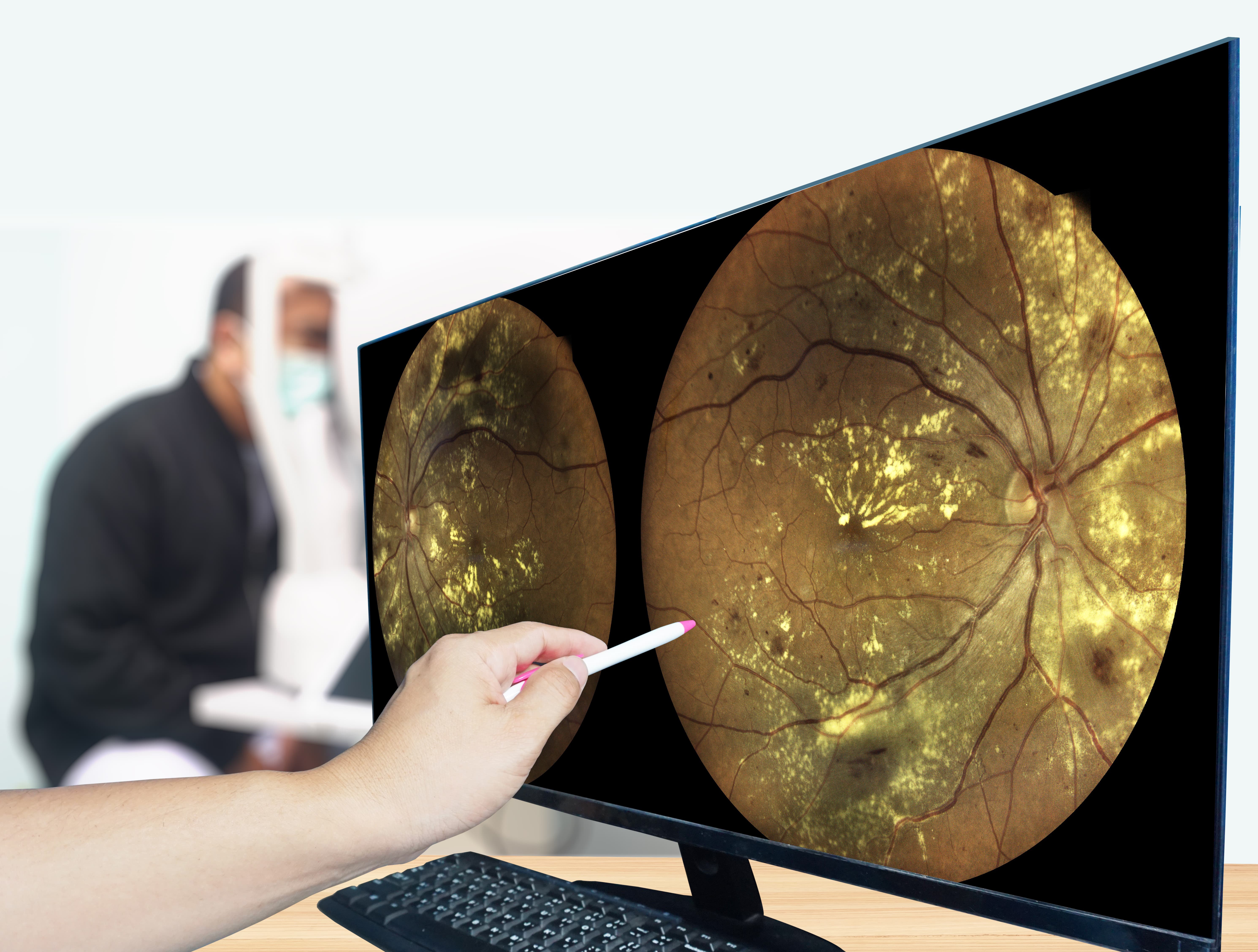
A complete structural and functional imaging record of your eyes, built so this year's findings can be measured against last year's.